3D Printed Anisotropic Tissue Simulants with Embedded Fluid Capsules Developed
Researchers have developed 3D printed anisotropic tissue simulants containing embedded fluid capsules, offering a new tool for medical simulation and training.
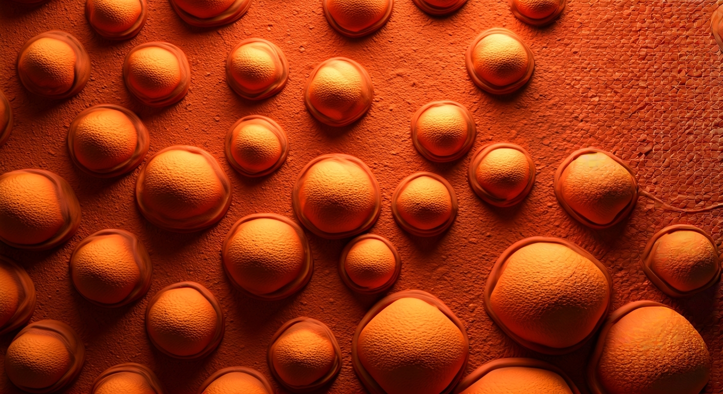
A novel method for creating 3D printed anisotropic tissue simulants has been reported, integrating embedded fluid capsules within the printed structures. This development aims to enhance the realism and effectiveness of medical simulation and training scenarios.
The anisotropic nature of the simulants means they exhibit directional properties, mimicking the complex, non-uniform structure of biological tissues. This characteristic is crucial for accurately replicating the feel and behavior of actual human organs and tissues during surgical practice or medical device testing.
The inclusion of fluid capsules within these simulants adds another layer of fidelity. These capsules can be designed to rupture or release fluids upon interaction, simulating bleeding or the release of bodily fluids. This feature provides trainees with dynamic feedback, allowing them to practice techniques for managing such situations in a controlled environment.
The researchers highlight that this technology can be used to create realistic models for a variety of medical procedures. The ability to customize the mechanical properties and the behavior of the embedded capsules allows for the simulation of diverse anatomical structures and pathological conditions. This advancement holds potential for improving surgical skills and diagnostic training.
This development addresses the need for more realistic and dynamic training tools in medicine. By creating anisotropic materials with embedded fluid capsules, researchers are enabling the simulation of complex tissue behaviors and fluid dynamics, crucial for advanced surgical training. This pushes the boundaries of bioprinting beyond static models, moving towards functional, responsive anatomical replicas.
Edited by the news editor with AI from the original report — please refer to the original source.